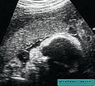
Although an ultrasound scan of the baby's heart is performed, echocardiography or fetal echocardiography It is a more specific test indicated for some special cases.
It is done to study the cardiac structure and functioning of the baby's heart before birth in the following cases.
When the ultrasound is suspected that there may be some cardiac abnormality, when there is a family history of heart disease, when there are diseases of the mother such as diabetes, rubella or alcoholism that could affect the functioning of the baby's heart.
Also when the mother has taken drugs or drugs that could cause cardiac malformations in the fetus or when there is a nuchal translucency greater than 4 millimeters,
It is a test that lasts about thirty minutes and is usually performed at week 20, although in some cases early echocardiography is used, around week 12 or 14.
The importance of carrying it out in the mother's womb is so that in the event that there is any problem, an operation can be performed soon after the baby is born.
In any case, the gynecologist will evaluate in each case if it is necessary to perform it. In my case, having a triple screenning of high risk, I also had an echocardiography to get out of doubt.